Research into two unstudied polyubiquitin chain types reveals complex nature of ubiquitin signals
Ubiquitylation regulates diverse cellular processes and this versatility is made possible because polyubiquitin chains of eight different linkage types can be assembled. Differently linked polyubiquitin chains couple to functionally distinct outcomes. For instance, proteins modified with K48-linked polyubiquitin are targeted for proteasomal degradation. Until now, nothing was known about K29 and K33 polyubiquitin chains and there weren't any methods to study them. Research carried out by Yosua Kristariyanto, a PhD student in Yogesh Kulathu's group, now provides exciting insights into two these two unstudied polyubiquitin modifications.
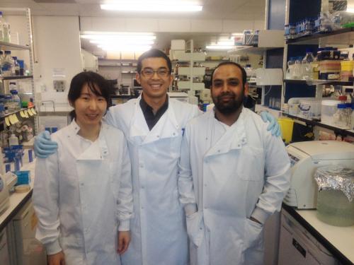
In two new papers just published in Molecular Cell and Biochemical Journal, Yosua together with Soo-Youn Choi have identified ubiquitin chain editing complexes, that combine E3 ligases with linkage selective deubiquitinases (DUBs), to enzymatically assemble K29 and K33 polyubiquitin chains. Importantly, these methods allow large-scale assembly using wild type ubiquitin yielding pure polyubiquitin chains, which will enable research into these uncharacterized modifications. For the first time, polyubiquitin tetramers of seven different linkage types could be assembled, which enabled Yosua to screen linkage selectivity of ubiquitin binding domains (UBDs). This led to the discovery of the NPL4-like zinc finger (NZF) domains of TRABID as selective binders to K29 and K33 chains.
To understand the structural basis of selective recognition, Yosua with the help of Syed Arif Abdul Rehman, a postdoc in Kulathu lab, solved crystal structures of K29-linked diubiquitin, K33-linked diubiquitin and triubiquitin, and the first NZF of TRABID (NZF1) in complex with K29-linked diubiquitin. Intriguingly, in the crystal structure of the complex, K29 chains adopt a helical filament-like structure and further studies are required to address if such filamentous structures are formed in cells and what roles they may have. The structure of the complex also explains how the tandem NZF domain repeats of TRABID may recognize long K29 and K33 chains. Through detailed biochemical and biophysical analyses, the mechanisms underlying polyubiquitin recognition by NZF domains are revealed and highlights how despite their small size, NZF domains have evolved distinct mechanisms to achieve linkage-selective polyubiquitin recognition.
Taking these findings further, Yosua then developed methods to study K29 and K33 polyubiquitin chains in cells. Using the newly discovered linkage-specific binders as affinity reagents reveals that K29 chains are made in cells as short polymers (2-4 ubiquitin moieties). Intriguingly these short K29 chains are found within polyubiquitin containing other linkages. This is an emerging theme in the ubiquitin field that heterotypic polyubiquitin chains containing multiple linkages can be formed and function as specialised signals. The heterotypic nature of polyubiquitin adds to the complexity of the already information rich ubiquitin system, and will require novel approaches to study them.
To read a copy of Yosua's papers that have just been published, click here (K29) and here (K33).

