


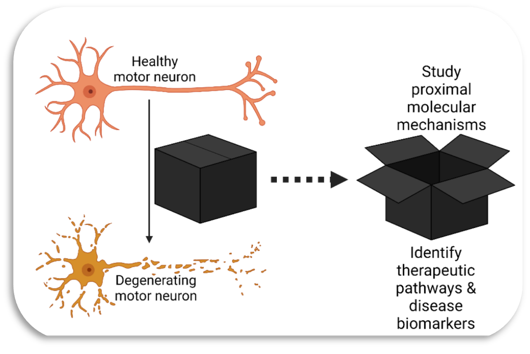
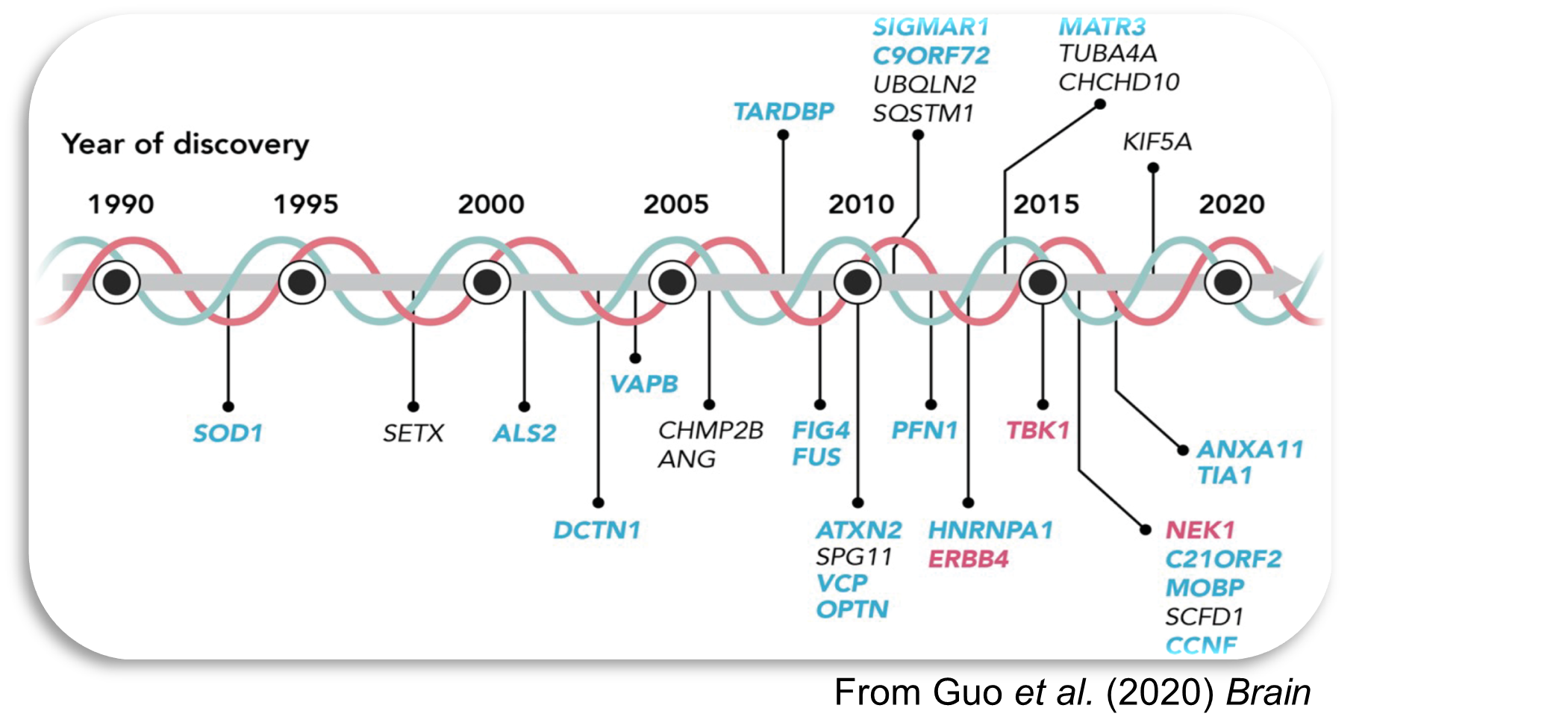
Our laboratory studies cell signalling in amyotrophic lateral sclerosis (ALS), a fatal neurological disorder characterised by the degeneration of motor neurons, for which there is still no effective disease-modifying treatment. Genetics has implicated a series of kinases and kinase-regulated proteins in ALS, among them NEK1 and TBK1, yet kinases have had comparatively little dedicated biochemical attention in the field.
Rare variants in NEK1 are found in 2–3% of people with ALS and have now been confirmed at exome-wide significance in the largest ALS sequencing study to date. Despite this, NEK1 itself remained a “dark kinase”: scarce inside cells, with no validated autophosphorylation site, no readout of its activity, and no antibodies or cell lines with which to work. Without those, a newly found variant could not be called damaging or harmless, and there was no rational way to drug the enzyme.
What we have shown
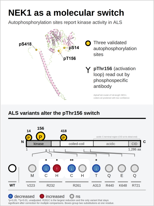
To address this, we generated a comprehensive cell-based phosphoproteomic map of NEK1, identifying ten recurrent phosphorylation sites and assigning three of them, pSer14, pThr156 and pSer418, as autophosphorylation sites, using kinase-dead controls, targeted extracted-ion chromatogram analysis, phosphosite mutagenesis and phosphospecific antibodies raised in-house.
Activation-loop pThr156 gives a quantitative readout of NEK1 activity. Applying it to nine ALS-associated missense variants spanning the major functional regions of the protein, we found that R261C caused the largest loss of autophosphorylation, R232C and A313T smaller reductions, and R232H an increase; structural modelling explains why each residue behaves as it does. This is the first activity-based framework for classifying NEK1 variants, and it shows directly that missense variants can alter NEK1 activity by a mechanism distinct from simple haploinsufficiency.
NEK1 does not work alone
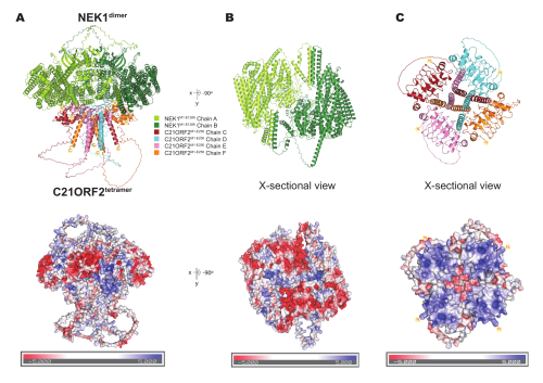
NEK1 forms a stable complex with C21ORF2, itself an ALS-associated ciliary and DNA-damage-response protein. Guided by our mass photometry data, we modelled a 2:4 NEK1–C21ORF2 assembly in which an acidic C-terminal face of the NEK1 dimer docks onto a basic N-terminal surface presented by four C21ORF2 chains. The model is a hypothesis-generating framework, rather than an experimentally determined structure, and testing it is a current priority.
Key questions for ongoing work
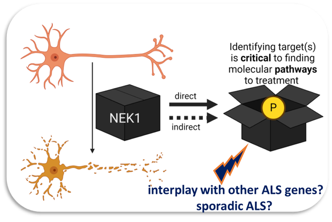
We are now asking what NEK1 phosphorylates in human motor neurons, and what those substrates do. Separately, a kinome-wide pooled CRISPR screen in C9orf72 motor neuron-microglia co-cultures, with Dr Wenting Guo in Strasbourg, will identify druggable modifiers of toxicity.
Arpan’s ambition, as a practising clinical neurologist, is to make fundamental discoveries, so that ALS is better understood, better diagnosed and eventually treated.
Joining the group
Arpan is currently applying for intermediate clinician-scientist fellowships to consolidate his work and enable him to build his own team. He is keen to hear from motivated postdoctoral researchers, PhD candidates and clinical trainees considering a research fellowship, who would like to work on kinase signalling in ALS. The laboratory sits in a Unit where all PIs share an ethos that prioritises personal wellbeing and career development, and a positive research culture. Informal enquiries are welcomed via email.
Publications
NEK1 and kinase signalling in ALS
- Agarwal S, Abdul Rehman SA, Muñoz IM, Knebel A, Hop PJ, Gourlay R, Brown F, Macartney T, Squires I, Veldink JH, Kenna KP, Rouse J, Mehta AR. NEK1 autophosphorylation is disrupted by amyotrophic lateral sclerosis-associated missense variants: activity biomarkers and structural insights. Open Biol. 2026 (accepted).
- Agarwal S, Gourlay R, Soares RF, Mehta AR. In-gel tryptic digestion and HCD/EThcD LC-MS/MS for mapping autophosphorylation sites. Bio-protocol. 2026 (in press).
ALS disease mechanisms in human stem cell models
- Mehta AR, Gregory JM, Dando O, Carter RN, Burr K, Nanda J, Story D, McDade K, Smith C, Morton NM, Mahad DJ, Hardingham GE, Chandran S, Selvaraj BT. Mitochondrial bioenergetic deficits in C9orf72 amyotrophic lateral sclerosis motor neurons cause dysfunctional axonal homeostasis. Acta Neuropathol. 2021;141:257–279.
- Banerjee P, Mehta AR, Nirujogi RS, Cooper J, James OG, Nanda J, Longden J, Burr K, McDade K, Salzinger A, Paza E, Newton J, Story D, Pal S, Smith C, Alessi DR, Selvaraj BT, Priller J, Chandran S. Cell-autonomous immune dysfunction driven by disrupted autophagy in C9orf72-ALS iPSC-derived microglia contributes to neurodegeneration. Sci Adv. 2023;9:eabq0651.
- Fumagalli L, Young FL, Boeynaems S, De Decker M, Mehta AR, et al., Van Damme P. C9orf72-derived arginine-containing dipeptide repeats associate with axonal transport machinery and impede microtubule-based motility. Sci Adv. 2021;7:eabg3013.
- Braems E, Bercier V, Van Schoor E, Heeren K, Beckers J, Fumagalli L, Dedeene L, Moisse M, Geudens I, Hersmus N, Mehta AR, et al., Van Den Bosch L. HNRNPK alleviates RNA toxicity by counteracting DNA damage in C9orf72 ALS. Acta Neuropathol. 2022;144:465–488.
- Zhao C, Devlin AC, Chouhan AK, Selvaraj BT, Stavrou M, Burr K, Brivio V, He X, Mehta AR, et al., Chandran S. Mutant C9orf72 human iPSC-derived astrocytes cause non-cell autonomous motor neuron pathophysiology. Glia. 2020;68:1046–1064.
- Mehta AR, Chandran S, Selvaraj BT. Assessment of mitochondrial trafficking as a surrogate for fast axonal transport in human induced pluripotent stem cell-derived spinal motor neurons. Methods Mol Biol. 2022;2431:311–322.
Clinical trials & trial innovation in ALS
- Pal S, Chataway J, Swingler R, Macleod MR, Carragher NO, Hardingham G, Selvaraj BT, Smith C, Wong C, Newton J, Lyle D, Stenson A, Dakin RS, Ihenacho A, Colville S, Mehta AR, et al., Chandran S; MND SMART Investigators. Safety and efficacy of memantine and trazodone versus placebo for motor neuron disease (MND-SMART): stage two interim analysis from the first cycle of a phase 3, multiarm, multistage, randomised, adaptive platform trial. Lancet Neurol. 2024;23:1097–1107.
- Mehta AR, Carpenter JR, Nicholas JM, Chataway J, Virgo B, Parmar MKB, Chandran S, Pal S. The role of placebo control in clinical trials for neurodegenerative diseases. Nat Med. 2023;29:2682–2683.
- Mehta AR, Pal S, Chataway J, Carpenter JR, Parmar MKB, Chandran S. Smarter adaptive platform clinical trials in neurology: a showcase for UK innovation. Brain. 2022;145:e64–e65.
- Mehta AR, Chataway J, Pal S, Parmar MKB, Chandran S. Trials for neurodegenerative diseases: time to innovate. Lancet Neurol. 2021;20:984.
ALS genetics, epidemiology and care
- Mehta PR, Iacoangeli A, Opie-Martin S, van Vugt JJFA, Al Khleifat A, Bredin A, Ossher L, Andersen PM, Hardiman O, Mehta AR, Fratta P, Talbot K; Project MinE ALS Sequencing Consortium; Al-Chalabi A. The impact of age on genetic testing decisions in amyotrophic lateral sclerosis. Brain. 2022;145:4440–4447.
- Glasmacher SA, Kearns PKA, Larraz J, Stirland L, Mehta AR, Newton J, Weir CJ, Chandran S, Pal S; CARE-MND Consortium. Prevalence of multimorbidity and its impact on survival in people with motor neuron disease. Eur J Neurol. 2021;28:2756–2765.
- Ng Kee Kwong KC, Gregory JM, Pal S, Chandran S, Mehta AR. Cerebrospinal fluid cytotoxicity in amyotrophic lateral sclerosis: a systematic review of in vitro studies. Brain Commun. 2020;2:fcaa121.
- Mehta AR, Selvaraj BT, Barton SK, McDade K, Abrahams S, Chandran S, Smith C, Gregory JM. Improved detection of RNA foci in C9orf72 amyotrophic lateral sclerosis post-mortem tissue using BaseScope shows a lack of association with cognitive dysfunction. Brain Commun. 2020;2:fcaa009.
- Jayaprakash K, Glasmacher SA, Pang B, Beswick E, Mehta AR, Dakin R, Newton J, Chandran S, Pal S; CARE-MND Consortium. Riluzole prescribing, uptake and treatment discontinuation in people with amyotrophic lateral sclerosis in Scotland. J Neurol. 2020;267:2459–2461.
Clinical neurology, global neurology & the culture of neuroscience
- Sudarshan R, Sayo AR, Renner DR, de Saram S, Godbole G, Warrell C, Duong HTH, Thwaites CL, Mehta AR*, Coughlan C*. Tetanus: recognition and management. Lancet Infect Dis. 2025;25:e645–e657.
- Coughlan C, Sudarshan R, Mehta AR. What comes next for global neurology? Brain. 2026;149:2569–2573.
- Devine H, Jabbari E, Scott J, Mehta AR, Dobson R, Mead S. Academic neurology in the UK: a plea to turn away from the precipice. Brain. 2024;147:2270–2273.
- Takahashi K, Agarwal S, Mehta AR. 20 years of iPS cells. Lancet Neurol. 2026;25:631.
- Mehta AR, Mehta PR, Anderson SP, MacKinnon BLH, Compston A. Grey Matter: Etymology and the neuron(e). Brain. 2020;143:374–379.
- Mehta AR, Abbott CM, Chandran S, Haley J. The Cajal Embroidery Project: celebrating neuroscience. Lancet Neurol. 2020;19:979.
* denotes joint senior authorship.
Open science and reagents
Raw mass spectrometry data from the Open Biology paper are deposited in PRIDE (PXD079815) and the analysis files in Zenodo; the preprint is on bioRxiv. The reagents are available to any laboratory that asks. Both of the group’s principal methods are published as open protocols (Methods in Molecular Biology, 2022; Bio-protocol, 2026).
Collaborators
Professor Jan Veldink, Department of Neurology, UMC Utrecht Brain Center, University Medical Center Utrecht, Utrecht University, The Netherlands.
Dr Kevin Kenna, Department of Translational Neuroscience, UMC Utrecht Brain Center, University Medical Center Utrecht, Utrecht University, The Netherlands.
Professor Kevin Talbot, Head of Department & Professor of Motor Neuron Biology, University of Oxford.
Professor John Rouse, Professor of Chromosome Biology, Division of Genome Integrity, Faculty of Life Sciences, University of Dundee.
Professor Colin Smith, Director of Centre for Clinical Brain Sciences & Professor of Neuropathology, Euan MacDonald Centre for MND Research, University of Edinburgh.
Dr William Farnaby, Principal Investigator, Centre for Targeted Protein Degradation, Faculty of Life Sciences, University of Dundee.
Dr Wenting Guo, Principal Investigator, NeuroStra Institute, Strasbourg, France.
Dr Raja Nirujogi, Independent Investigator, MRC PPU, Faculty of Life Sciences, University of Dundee.