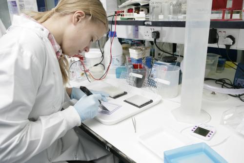
Agne Kazlauskaite defines PINK1 as the first ubiquitin kinase
Exactly 10 years ago mutations in a little studied kinase called PTEN-induced kinase 1 (PINK1) were discovered in patients with early-onset Parkinson's disease [Ref 1]. PINK1 is unique amongst all kinases in possessing a N-terminal targeting domain that localizes it to the mitochondria. There has been great interest in understanding the function of PINK1 however, the human enzyme was difficult to express and exhibited very low levels of kinase activity, which hampered early progress.
In 2011, PhD student, Helen Woodroof, (supervised by Miratul Muqit, Daan van Aalten and Dario Alessi) made a breakthrough by discovering constitutively active insect orthologues of PINK1, which led to the establishment of the first assays of PINK1 kinase activity in the field [Ref 2]. Another PhD student, Chandana Kondapalli (supervised by Miratul Muqit and Dario Alessi) then deployed insect PINK1 in a screen to identify the Parkinson's linked ubiquitin E3 ligase, Parkin as the first PINK1 substrate. Chandana mapped the phosphorylation site to Serine65 (Ser65) that lies within the N-terminal Ubiquitin-like (Ubl) domain of Parkin [Ref 3]. She also raised a phospho-specific antibody to demonstrate that the Parkin Ser65 site was phosphorylated by PINK1 in cells in vivo.
Parkin is a RING-IBR-RING E3 ubiquitin ligase and mutations had been discovered in Parkinson's patients in 1998. For many years it was believed that Parkin was constitutively active. However, Helen Walden whilst at the CRUK laboratories in Lincolns Inn Fields, made a major advance by discovering that Parkin is autoinhibited and showed that this was mediated by its Ubl domain [Ref 4]. However, the physiological signal to convert Parkin from an inactive to active conformation was unknown. Agne Kazlauskaite, a PhD student (supervised by Dario Alessi and Miratul Muqit) set out to test whether PINK1-dependent phosphorylation of Parkin at Ser65 could be that signal and in a series of experiments published in two papers demonstrated that Parkin Ser65 phosphorylation indeed was critical for Parkin activation as judged by monitoring the ubiquitylation of the Parkin substrate Miro1 as well as Parkin autoubiquitylation and the formation of free polyubiquitin chains [Ref 3 and Ref 5].
However, an outstanding question was how PINK1-dependent phosphorylation of Parkin at Ser65 led to activation of its ubiquitin E3 ligase activity. The answer came unexpectedly when Chandana Kondapalli working with Matthias Trost's lab undertook a phosphoproteomic screen to identify novel PINK1 substrates. Strikingly they identified a novel ubiquitin phosphopeptide phosphorylated at Ser65 that was significantly enriched in cells over-expressing wild-type PINK1 activated by the mitochondrial-uncoupling agent, CCCP, compared to cells expressing kinase-inactive PINK1. Ser65 in ubiquitin lies in a similar motif to Ser65 in Parkin's Ubl domain.
Agne Kazlauskaite next investigated the functional role of ubiquitin Ser65 phosphorylation. Remarkably Agne found that PINK1 could directly phosphorylate ubiquitin at Ser65. In a series of experiments she next demonstrated that Ser65-phosphorylated ubiquitin (ubiquitinPhospho-Ser65) was an activator of Parkin. She initially demonstrated that wild-type but not kinase-inactive PINK1 could still activate a fragment of Parkin that lacks the Ubl domain (encompassing the Ser65 residue) (Ubl-Parkin). Axel Knebel's lab then expressed and purified ubiquitinPhospho-Ser65 and the isolated Parkin Ubl domain phosphorylated at Ser65 (UblPhospho-Ser65). Agne was able to show that ubiquitinPhospho-Ser65 but not non-phosphorylated ubiquitin could activate Ubl-deleted Parkin. She also found that UblPhospho-Ser65 could also activate this Ubl-deleted Parkin similar to ubiquitinPhospho-Ser65. Interestingly Agne found that ubiquitinPhospho-Ser65 could activate full-length Parkin but this was not the case for non-phosphorylated ubiquitin or UblPhospho-Ser65. Finally Agne was able to show that both PINK1-dependent phosphorylation of Parkin at Ser65 as well as ubiquitin at Ser65 is required for optimal activation of Parkin E3 ligase activity since only mutation of both proteins at Ser65 leads to complete loss of Parkin E3 ligase activity.
Agne's new findings suggest that small molecule activators of Parkin that mimic ubiquitinPhospho-Ser65 could represent a promising therapy for Parkinson's. Her work also provides the most stark example to date of the convergence of protein phosphorylation with protein ubiquitylation and opens a new area of research in determining the role of phospho-ubiquitin in Parkinson's disease and beyond.
To read a copy of Agne's paper published in Biochemical Journal click here

