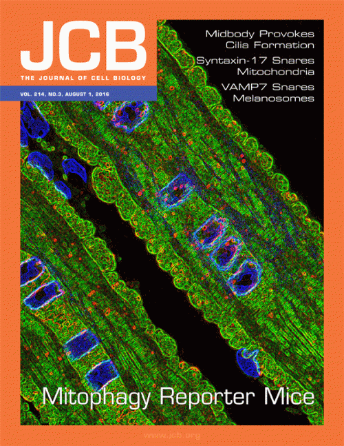
Mitochondria are the essential energy-generating powerhouses that provide our cells with the energy of life. However, their malfunction has a dark side. Damaged mitochondria have the potential to release destructive reactive oxygen species that have serious and deleterious consequences for the cell. To cope with this, our cells have evolved a protective mechanism to prevent such a “mitochondrial meltdown”, by eliminating damaged mitochondria through a process termed mitophagy. Mitophagy is a cellular waste disposal pathway that delivers faulty and superfluous mitochondria to lysosomes for degradation and recycling. Over the past decade, landmark studies in cultured cells have hinted that mitophagy may be a vital “stress response” that sustains the health of the mitochondrial network or “cellular power grid” when damaged. However, it has been very difficult to visualise this process in tissues and thus, little is known about the true nature of mitophagy in whole organisms. Several important questions remain unanswered - do our cells perform mitophagy all the time, or only under conditions of stress? Likewise, in what tissues and in what kind of cells within our organs does mitophagy proceed? It is vital our knowledge of this process extends beyond the Petri dish to a more biomedical context, especially as mitophagy may be critical in human disease progression. It has recently been proposed that impaired mitophagy may be instrumental in triggering neurodegeneration in some forms of hereditary Parkinson’s disease.
Pioneering research from the Ganley Lab has now shed light on the physiological instances of mitophagy. In a cover article published by the Journal of Cell Biology, research spearheaded by postdoctoral fellow Tom McWilliams describes a fluorescence-based mitophagy mouse model, which they have called mito-QC (for mitochondrial Quality Control). mito-QC allows the visualisation of mitochondrial morphology and turnover in defined cells within tissues for the first time. The work, which was a collaborative effort between the Ganley and Muqit Labs in the MRC-PPU as well as Alan Prescott and the SLS Dundee Imaging Facility, has really opened up the field of mitophagy. This work clearly shows that mitophagy is a physiological process and even in healthy animals, mitophagy is occurring in multiple cell types within diverse tissues. Interestingly, the amount of mitophagy appears to vary between specific cells within tissues. For example, proximal tubule cells within the adult kidney exhibit an extremely high level of mitophagy while the related distal tubule cells, in the same organ, do not. It is of note that impaired proximal tubule function is involved in renal failure and kidney disease and, given its protective function, it is possible that mitophagy plays an important role here. Likewise, Tom was able to show that mitophagy is happening in distinct neurons of the brain and by crossing mito-QC with multiple mouse models of Parkinson’s disease, it is the hope that mitophagy can be finally studied in brain regions that are relevant to disease pathology. Knowing when and where this pathway is disrupted may provide critical knowledge in developing treatments for this currently incurable disease, as well as for other diseases where mitophagy has been implicated.
A major aim will now be to determine the molecular cues that trigger and regulate mitophagy in tissues, in order to understand how disrupting this quality control pathway could lead to disease onset and progression.

