

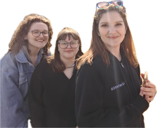
Two PhD students from the Madsen Lab at the MRC Protein Phosphorylation and Ubiquitylation Unit (MRC PPU), Oliwia Mruk and Alexandra Musk, have developed optimised workflows for generating arterial- and venous-like endothelial cells from human induced pluripotent stem cells (iPSCs). Their complementary 2D- and 3D-based workflows will be published in the September 2026 issue of STAR Protocols.
The research was initiated by Dr Ralitsa Madsen when she established her laboratory at the MRC PPU. It was motivated by the need for better human models of vascular malformations caused by mutations in PIK3CA, a gene encoding a key component of the phosphoinositide 3-kinase (PI3K) signalling pathway.
Existing methods for differentiating iPSCs into arterial- and venous-like endothelial cells relied on high concentrations of a PI3K inhibitor. This presented a fundamental problem: the inhibitor would also suppress the enzyme encoded by PIK3CA, making the resulting cells unsuitable for studying diseases driven by mutations in this gene.
Dr Madsen recognised that the original protocol also used high concentrations of insulin, a hormone that potently activates PI3K signalling. This insight enabled the team to remove both insulin and the PI3K inhibitor at key stages of arteriovenous differentiation. The result was a robust workflow compatible with modelling PIK3CA-driven vascular disease.
First author Oliwia Mruk played an instrumental role in benchmarking the revised workflow. Her work included extensive characterisation of endothelial-cell maturation during continuous exposure to unidirectional shear stress – mimicking the mechanical force created by blood flow – to better reproduce the physiological conditions experienced by endothelial cells in the body.
Second author Alex Musk established a complementary 3D differentiation system. She adapted Dr Madsen’s existing high-throughput 3D culture workflow for quantitative, single-cell studies of signalling using mass cytometry (Madsen et al. 2025 Mol Sys Bio). Unexpectedly, differentiation in 3D substantially increased the efficiency with which venous-like endothelial cells could be generated. This improvement will allow the team and other researchers to produce this disease-relevant cell type in the numbers required for robust mechanistic studies.
Together, the protocols provide high-fidelity human endothelial-cell models for investigating how genetic activation of PI3K signalling disrupts vascular development. They also offer a foundation for evaluating potential therapeutic strategies in experimentally controlled, disease-relevant systems.
The work was supported by CLOVES Syndrome Community (CSC), a UKRI Future Leaders Fellowship awarded to Dr Madsen, an MRC PhD studentship awarded to Oliwia Mruk, and a Wellcome Trust PhD studentship awarded to Alex Musk.
Following publication, CSC Director Lauren Beauregaard said:
“We at CSC look forward to continued collaboration with Dr Ralitsa Madsen and her team. We trust their science, their ethics, and their intention, and know that through their work, people with CLOVES Syndrome and other PIK3CA-related disorders have brighter futures on the horizon.”
For Alex and Oliwia, the papers provided their first experience of taking research manuscripts through peer review, revision and proof production. They will now apply the models in their respective PhD projects. Oliwia is investigating non-cell-autonomous PI3K signalling – how genetically altered cells influence surrounding cells – while Alex is developing lineage- and mutation-resolved models of genetic PI3K pathway activation.