2024 represents the 20th anniversary since the first report of autosomal recessive mutations in the PINK1 kinase in Parkinson’s disease by the lab of Nick Wood. Since then pioneering studies by Richard Youle’s lab have identified that PINK1 together with another Parkinson’s gene Parkin function in a common pathway that removes damaged mitochondria by autophagy (mitophagy). Previous work by the Muqit lab and others found that PINK1 is activated upon mitochondrial damage to phosphorylate Ubiquitin and Parkin at an equivalent Serine 65 residue leading to activation of Parkin E3 ligase activity, a critical step for induction of mitophagy. Until recently how PINK1 is activated by mitochondrial stress was less understood.
In 2012 Mike Lazarou working in the Youle lab discovered that active PINK1 is stabilized at the Translocase of outer membrane (TOM) complex that resides on the outer mitochondrial membrane. The TOM complex comprises seven subunits, 5, 6, 7, 20, 22, 40 and 70 and further work from the Youle lab established a critical role of the TOM7 subunit for PINK1 activation. In 2022 the Muqit lab discovered an intramolecular interface formed by an N-terminal a-helix extension (NTE) and C-terminal a-helix extension (CTE) of PINK1 that lies outside the kinase domain. Critically PD-associated mutations located at the NTE:CTE interface disrupted PINK1 recruitment to the TOM complex and activation. This was also independently found by the labs of David Komander and Jean-Francois Trempe.
Further understanding of how PINK1 is activated at the TOM complex has now been solved in new work published in Science Advances, by the Muqit Lab in collaboration with the Labib & Kulathu labs in MRC-PPU and researchers from the University of Leeds, University Medical Center Göttingen, and Goethe-Universität, Frankfurt.
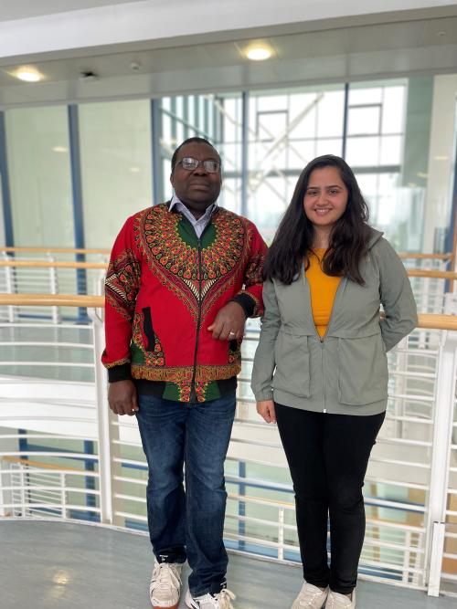
The study was led by post-docs Olawale (Wale) Raimi and Hina Ojha. Wale working with Cristian Polo Rivera and Tom Deegan in Karim Labib’s lab developed a method to express human PINK1 and all 7 subunits of the human TOM complex in budding yeast. Yeast do not express PINK1 but Kenneth Ehses, a PhD student in the lab of Rubén Fernández Busnadiego, confirmed that the human PINK1-TOM complex was indeed localized on yeast mitochondria. Strikingly Wale discovered that co-expression of human PINK1 and TOM subunits led to PINK1 activation. He then employed genetic analysis in yeast to dissect the contribution of individual TOM subunits towards PINK1 activation revealing a critical role for the pore-containing subunit TOM40 and supporting subunits TOM7 and TOM22. Further he found an important role for the accessory subunits TOM20 and TOM70.
Hina working with Sven Lange in Yogesh Kulathu’s lab developed a high confidence structural model of the PINK1-TOM complex using AlphaFold2. This revealed that the PINK1 NTE:CTE interface binds TOM20 and she also was able to identify a region located in the N-terminus of PINK1 that binds TOM70 termed the TOM70-interacting region (TIR). Hina undertook extensive mutagenesis analysis in human mammalian cells exposed to mitochondrial damage to verify the predicted model and PINK1-TOM interfaces and this work required much molecular biology support from Mel Wightman and Rachel Toth from the MRC-PPU cloning team.
Overall, these findings will stimulate further ideas on how to activate PINK1 as a potential strategy for treating Parkinson’s patients.
The research was funded by the Wellcome Trust, Aligning Science Across Parkinson’s (ASAP), Michael J. Fox Foundation, and the Medical Research Council.
To read a copy of the paper published in Science Advances click here https://www.science.org/doi/epdf/10.1126/sciadv.adn7191

