In 1998, mutations in the Parkin gene were discovered as a cause of early-onset Parkinson’s disease by a Japanese team who in collaboration with Keiji Tanaka then showed in 2000 that it functioned as a ubiquitin E3 ligase. However, the physiological substrates of Parkin remained elusive for many years. A major breakthrough on Parkin’s function was the discovery, by Richard Youle’s lab, that Parkin is recruited from the cytosol to sites of mitochondrial damage to trigger the removal of mitochondria by autophagy known as mitophagy.
In parallel work, research in the laboratory of Miratul Muqit discovered that upon mitochondrial damage, another Parkinson’s linked gene PINK1 can phosphorylate Parkin at a highly conserved residue, Serine 65 (Ser65), that lies within its N-terminal Ubiquitin-like domain (Ubl). Further, his group and others discovered that PINK1 can phosphorylate Ubiquitin at an equivalent Ser65 residue and this associates with Parkin in a complex to trigger maximal activation of Parkin E3 ligase activity.
The laboratory of Wade Harper at Harvard Medical School has pioneered the development of proteomic detection and mapping of ubiquitylation sites in complex cell extracts. Spearheaded by a post-doc Alban Ordureau (who is a former PhD student of Philip Cohen), the Harper lab has mapped many Parkin substrates at the mitochondrial outer membrane of cells and this work has contributed to general understanding of the early signalling required for mitophagy.
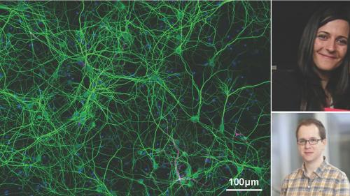
However, to date very little was known about Parkin substrates in cell types relevant to Parkinson’s disease including neurons, and under conditions in which Parkin and PINK1 are expressed under endogenous levels. To address this question, PhD student, Odetta Antico, built on previous work by post-doc Erica Barini to establish a robust system to monitor the PINK1 and Parkin pathway biochemically from isolated mouse cortical neurons. This included the observation that PINK1-dependent phosphorylation of Rab proteins is Parkin-independent in conjunction with a visiting undergraduate student, Mollie Rickwood. In complementary analysis Francois Singh from Ian Ganley’s lab performed metabolic profiling of wild-type and PINK1 knockout neurons and ASAP-funded senior post-doc, Raja Nirujogi, undertook mass spectrometry-based copy number analysis to define the precise levels of Parkin and other components in the isolated neurons.
Odetta then teamed up with Alban in the Harper lab to perform ubiquitylation analysis of post-mitotic cortical neurons derived from wild-type mice. Their analysis revealed nearly 10,000 diGLY-containing ubiquitylated (Kgg) peptides in neurons of which 160 Kgg peptides from 70 mitochondrial proteins were significantly elevated upon mitochondrial depolarisation induced by AntimycinA/Oligomycin. They next undertook similar analysis in wild-type and PINK1 knockout neurons and this identified sites that were PINK1-dependent. Strikingly comparative analysis of PINK1-regulated sites in mouse neurons with data obtained previously by Alban from human iNeurons (that share similarities to cortical neurons) pinpointed only 49 Kgg sites across 22 proteins that were conserved across mouse and human neurons. These proteins included CISD1, CPT1A, CYB5B, HK1, MFN2and VDAC1.
To validate that these sites were indeed PINK1 and Parkin regulated, Odetta performed a variety of cell-based assays in PINK1 and Parkin knockout neurons including ubiquitin capture experiments combined with western blotting and reconstitution experiments in isolated mitochondria. Furthermore, Michael Stevens, a post-doc in the Muqit lab, performed in vitro assays with purified protein to demonstrate direct modification of substrates by Parkin (pre-activated with PINK1).
Overall the findings of Odetta, Alban and colleagues has furthered general understanding of how these 2 Parkinson’s proteins, PINK1 and Parkin regulate mitophagy which is conserved in nearly every cell type including neurons.
Furthermore, their results will aid in the development of novel biomarkers and small molecular activators of PINK1 and/or Parkin as a potential therapy against Parkinson’s.
The research was funded by the Wellcome Trust, Medical Research Council, Parkinson’s UK, Rosetrees Trust, Michael J Fox Foundation, EMBO, Dundee Research Interest Group (DRIG), NIH, a gift from N. Goodnow (to Wade Harper), Bath University Alumni Placement Grant, and Aligning Science Across Parkinson’s (ASAP) initiative.
To read a copy of the paper published in Science Advances click here.
The Kgg sites can be searched on a database termed MitoNUb click here.

